It all begins with a diagnosis
Gingivitis and periodontitis almost never involve pain. Over a period of years, the infection unobtrusively breaks down the supporting tissue around the teeth. Sometimes treatment comes too late, resulting in patients losing several or all of their remaining teeth.
What is your periodontal health like?
After an examination, a dentist or periodontologist can give you a clear picture of the tissue surrounding your teeth.
- He or she can detect any inflammation using a special measuring instrument (pocket periodontal probe). This pocket probe is placed between the tooth and the gum. Any bleeding or discharge of pus indicates an infection. Deepened pockets of more than 4 mm are usually a sign of jawbone loss and hence periodontitis.
- The clinical examination is normally followed by and X-ray examination, adjusted to the patient. A series of small intra-oral X-rays or a panoramic X-ray provide an image of the bone level around the teeth.
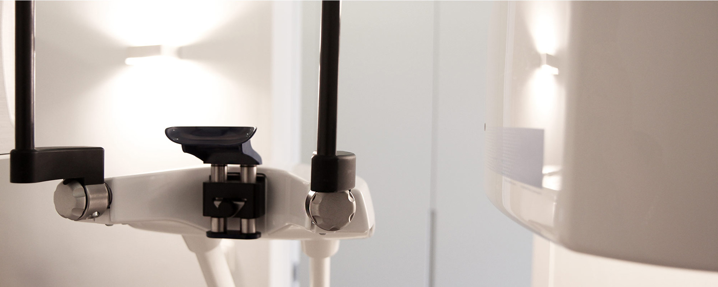
- Since 2007, we have been one of the first detal offices in the Benelux to take 3D images using a Cone Beam CT. A single imaging session lasting for less than 20 seconds can visualise both the upper and lower jaw. This process provides us with additional diagnostic information with only limited exposure to radiation for the patient.
